Boca Raton's First Choice For
Eye Care
Eye Exams
Eyeglasses
Contact Lenses
Watch Video
Modal Title
We Accept Most Insurance Plans
Highest Rated Eye Doctor in Boca
Prescription Eyewear In 1-Hour
We Accept Most Insurance Plans
Highest Rated Eye Doctor in Boca
Prescription Eyewear In 1-Hour
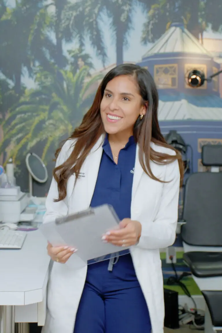
We're glad you found us
Going beyond the standard of eye care.
At Bocaview Optical, we go beyond the standard of eye care to ensure you receive the highest quality care. Whether you need an eye exam using the latest iWellness Technology, new glasses, contacts or eye products, we have you covered.
Our Patients Love Bocaview
We’ve been helping patients for over 30 years and have the highest rated google reviews in Boca Raton, Florida.
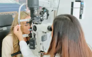
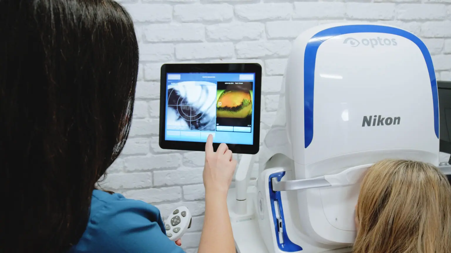
The Most Accurate Eye Exam
This is one of the few places in South Florida to offer the latest iWellness Exam and Keratograph 5M scanning technology.
Your Eye Health Matters To Us
Dr. Beck and our team of eye care specialists will help ensure your eyes remain healthy for years to come.
We have the largest selection of frames with over 1,000 top of the line frames in stock. You can now get your favorite brand sunglasses in your prescription.